Laboratory of X-Ray spectroscopy
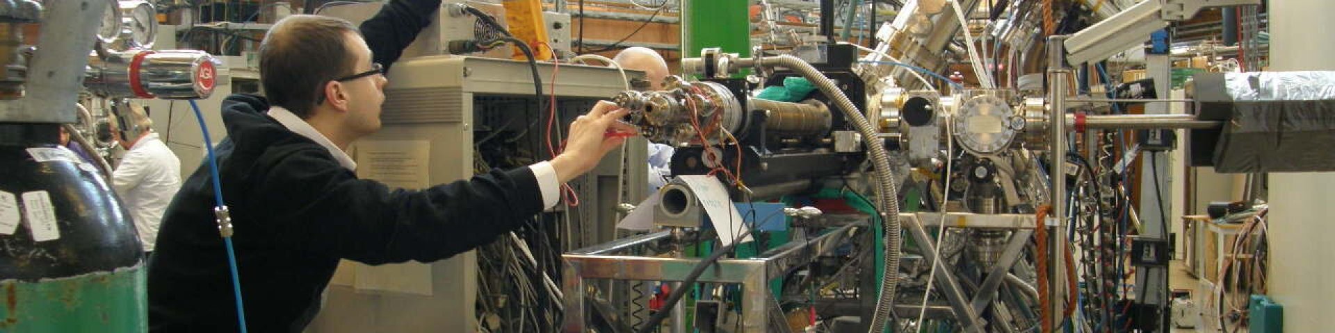
In laboratory of X-ray spectroscopy we use the soft X-ray excited processes for investigation of the atoms, molecules, thin films, solids and liquids. We also study antimicrobial surface coatings from an applied science perspective. We study the
- electronic structure of solid materials, thin films, nanoparticles and liquids;
- doping effects in thin oxide films;
- fragmentation of molecules under VUV irradiation;
- ionic liquids and in situ electrochemical processes;
- relaxation and decay processes of soft X-ray created excitations in solids.
For these studies we use: - X-ray photoabsorption spectroscopy;
- photoelectron spectroscopy;
- Auger spectroscopy;
- X-ray emission spectroscopy;
- time-of-flight mass spectroscopy;
- photoelectron-photoion coincidence spectroscopies.
- theoretical calculation methods (DFT, MP2, GW etc.)
For these studies we use synchrotron radiation at different beamlines of synchrotron radiation sources (MAX-IV in Lund, BESSY in Berlin, DESY in Hamburg). Together with three Finnish Universities we have built the FINEST branchline of beamline I3 at MAX-3 storage ring in MAX-lab for studies in VUV region. Also we have built the surface science station in our home lab, which basic instrument is photoelectron energy analyser Scienta SES-100. Our present great challenge is the development of the new beamline for soft X-ray studies at MAXlab’s new MAX-4 storage complex.
MAX-lab together with three Finnish Universities we have built the FINEST branchline of beamline I3 at MAX-3 storage ring in MAXlab for studies in VUV region. Also we have built the surface science station in our home lab, which basic instrument is photoelectron energy analyser Scienta SES-100. This equipment also consist of LHe crystate, SEM with resolution 1 um, LEED, time resolved luminescence equipment and sample preparation chamber with three electron gun evaporators. Our present great challenge is the development of the new beamline for soft X-ray studies at MAXlab’s new MAX-4 storage complex.
In collaboration with prof. Angela Ivask group (institute of molecular and cell biology, Tartu University) we study and develop antimicrobial surfaces. Microorganisms on various surfaces can enhance the spread of infections. Due to the increasing amount of antibiotic resistant bacteria there is strong interest among scientists and private enterprises of using antimicrobial surface coatings to prevent their spread.
|
Image
| Vambola Kisand Head of Lab Associate professor E-post: vambola.kisand@ut.ee Kabinet: B214 Publikatsioonid: Google Scholar |
|
Image
| Arvo Kikas Associate professor E-post: arvo.kikas@ut.ee Kabinet: B211 Publikatsioonid: Google Scholar |
|
Image
| Rainer Pärna Associate professor E-post: rainer.parna@ut.ee Kabinet: B212 Publikatsioonid: Google Scholar |
|
Image

| Tanel Käämbre Associate professor E-post: tanel.kaambre@ut.ee Kabinet: B212 Publikatsioonid: Google Scholar |
|
Image
| Arvo Tõnisoo engineer E-post: arvo.tonisoo@ut.ee Kabinet: B209 Publikatsioonid: Google Scholar |
|
Image
| Juhan Matthias Kahk Associate professor E-post: matthiaskahk@gmail.com Kabinet: B212 Publikatsioonid: Google Scholar |
|
Image
| Ivar Kuusik researcher E-post: ivar.kuusik@ut.ee Kabinet: B209 Publikatsioonid: Google Scholar |
|
Image

| Marta Berholts researcher E-post: marta.berholts@ut.ee Kabinet: B208 Publikatsioonid: Google Scholar |
|
Image

| Mati Kook Ph.D student E-post: mati.kook@ut.ee Kabinet: B210 Publikatsioonid: Google Scholar |
|
Image
| Dmytro Danilian researcher Kabinet: B208 |
|
Image
| Alexandra Nefedova laborant D308 |
|
Image

| Uday Pratap Singh Kushwah B208 Ph.D student |
|
Image

| Aadil Shafi Bhat B212 Ph.D student |
Core Electron Binding Energies in Solids from Periodic All-Electron Δ-Self-Consistent-Field Calculations
J. Matthias Kahk, Georg S. Michelitsch, Reinhard J. Maurer, Karsten Reuter, and Johannes Lischner
J. Phys. Chem. Lett. 2021, 12, 38, 9353–9359
Accurate absolute core-electron binding energies of molecules, solids, and surfaces from first-principles calculations
J. Matthias Kahk and Johannes Lischner
Phys. Rev. Materials 3, 100801(R)
Layer-resolved many-electron interactions in delafossite PdCoO2 from standing-wave photoemission spectroscopy
Lu, Q., Martins, H., Kahk, J.M., Rimal, G., Oh, S., Vishik, I., Brahlek, M., Chueh, W.C., Lischner, J. and Nemsak, S.
Communications Physics 4, no. 1 (2021): 1-8
Ionic Liquid Vapors in Vacuum: Possibility to Derive Anodic Stabilities from DFT and UPS
Kuusik, I., Kook, M., Pärna, R., Kisand, V.
ACS Omega, 2021
Frontier orbitals and quasiparticle energy levels in ionic liquids
Kahk, J.M., Kuusik, I., Kisand, V., Lovelock, K.R.J., Lischner, J.
npj Computational Materials, 2020, 6(1), 148
Selective antibiofilm properties and biocompatibility of nano-ZnO and nano-ZnO/Ag coated surfaces
Rosenberg, M., Visnapuu, M., Vija, H., ...Kahru, A., Ivask, A.
Scientific Reports, 2020, 10(1), 13478
UVA-induced antimicrobial activity of ZnO/Ag nanocomposite covered surfaces
Visnapuu, M., Rosenberg, M., Truska, E., ...Kisand, V., Ivask, A.
Colloids and Surfaces B: Biointerfaces, 2018, 169, pp. 222–232
UPS and DFT investigation of the electronic structure of gas-phase trimesic acid
Reisberg, L., Pärna, R., Kikas, A., ...Valden, M., Nõmmiste, E.
Journal of Electron Spectroscopy and Related Phenomena, 2016, 213, pp. 11–16
Co doped ZnO nanowires as visible light photocatalysts
Šutka, A., Käämbre, T., Pärna, R., ...Joost, U., Kisand, V.
Solid State Sciences, 2016, 56, pp. 54–62
Photocatalytic antibacterial activity of nano-TiO2 (anatase)-based thin films: Effects on Escherichia coli cells and fatty acids
Joost, U., Juganson, K., Visnapuu, M., ...Kisand, V., Ivask, A.
Journal of Photochemistry and Photobiology B: Biology, 2015, 142, pp. 178–185
Size-dependent toxicity of silver nanoparticles to bacteria, yeast, algae, crustaceans and mammalian cells in vitro
Ivask, A., Kurvet, I., Kasemets, K., ...Kisand, V., Kahru, A.
PLoS ONE, 2014, 9(7), e102108
Effect of cobalt doping and annealing on properties of titania thin films prepared by sol-gel process
Pärna, R., Joost, U., Nõmmiste, E., ...Kink, I., Kisand, V.
Applied Surface Science, 2011, 257(15), pp. 6897–6907
Resonant inelastic x-ray scattering and UV-VUV luminescence at the Be 1s edge in BeO
Kikas, A., Käämbre, T., Kooser, K., Pustovarov, V., Martinson, I.
Journal of Physics Condensed Matter, 2010, 22(37), 375505
Fragmentation and electronic decay of vacuum-ultraviolet-excited resonant states of molecular CsCl
Kisand, V., Kukk, E., Huttula, M., ...Nõmmiste, E., Aksela, S.
Journal of Physics B: Atomic, Molecular and Optical Physics, 2003, 36(19), pp. 3909–3921
Collapse of vibrational structure in the auger resonant raman spectrum of CO by frequency detuning
Sundin, S., mukhanov, F.K.G., Ågren, H., ...Ausmees, A., Svensson, S.
Physical Review Letters, 1997, 79(8), pp. 1451–1454
Autoionization phenomena involving the 2p53d configuration of argonlike ions in ionic solids
Elango, M., Ausmees, A., Kikas, A., ...Nyholm, R., Martinson, I.
Physical Review B, 1993, 47(18), pp. 11736–11748
The quantum yield spectra of electron emission of solids in XUV region
Elango, M., Kikas, A., Nǒmmiste, E., Pruulmann, J., Saar, A.
physica status solidi (b), 1982, 114(2), pp. 487–493
Photofragmentation of dual-function radiosensitizers
One of the leading strategies for cancer treatment is radiation therapy during which tumor cells are irradiated with highly energetic x-rays. The disadvantage of that treatment is that both cancerous and healthy cells located in the irradiated area will be damaged. Therefore, it is crucial to find a balance between inhibiting tumor growth and minimizing negative effects on the human body by controlling the dose of radiation. One solution is to use radiosensitizers, agents that enhance the damage to the cancerous cells. In this project, radiosensitizers based on nitroimidazole molecules will be enhanced with an iodine atom that is expected to act as an antenna that would help to localize the x-rays only in the tumor region avoiding major damage to the healthy cells. Understanding the ionization-induced reactions of radiosensitizers on the molecular level is crucial in evaluating their potential and in developing new and more effective drugs. In this project, mass spectrometric methods combined with synchrotron radiation will be used to reveal fragmentation pathways and the exact species formed upon irradiation of selected iodinated nitroimidazoles. Research results contribute to attempts to develop better radiosensitizers for radiation therapy.
List of research projects with participation from RSL emplyees:
Development of antimicrobial surface coatings tested in real-life conditions into pilot production phase
The X-Ray spektroscopy lab was founded by Mart Elango (1936-1996). After Mart the lab was headed by Arvo Kikas. From 1999 onwards, the professor and member of the Academy of sciences Ergo Nõmmiste was the scientific leader of the lab. Ergo passed away in 2019, after which the lab is headed by Dr. Vambola Kisand.
|
Image
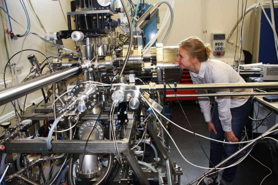
| X-ray photoelectron spectroscopy set-up (XPS)
Scienta SES-100 electron energy analyzer Nonmonochromatized Thermo XR3E2 twin anode X-ray source Al anode (photon energy 1486.6 eV) and Mg anode (photon energy 1253.6 eV).
|
|
Image
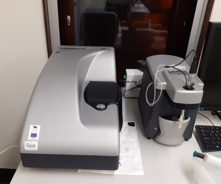
| Nanoparticle size analyzer Model: Malvern Zetasizer Nano ZSP Measurements of nanoparticle size in liquids with DLS method
|
|
Image
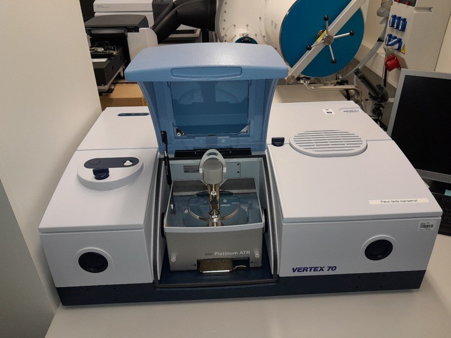
| Infrared FTIR spectrometer Vertex 70 Spectral range: 10 000 …. 50 1/cm |
|
Image
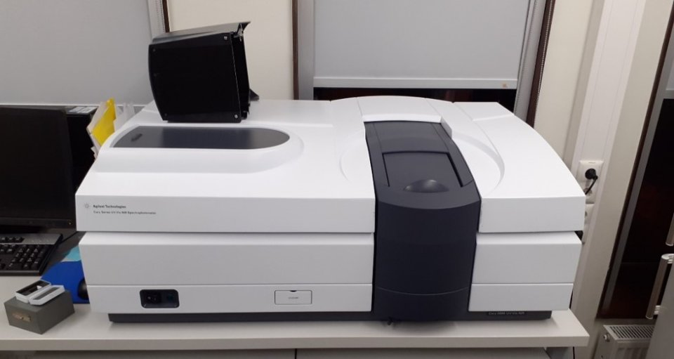
| UV-VIS-NIR spektometer Cary 5000 UV/VIS spectral region 190-900 nm
|
|
Image

| Environmental test chamber Memmert CTC 256 Volume 256 litres
|